


Radiographic
Oral Control
Radiographic Oral Examination
What is X-ray oral examination?
This test has a decisive role in the diagnosis and treatment dental problems that may not be visible to the naked eye. But how do oral exams actually work?
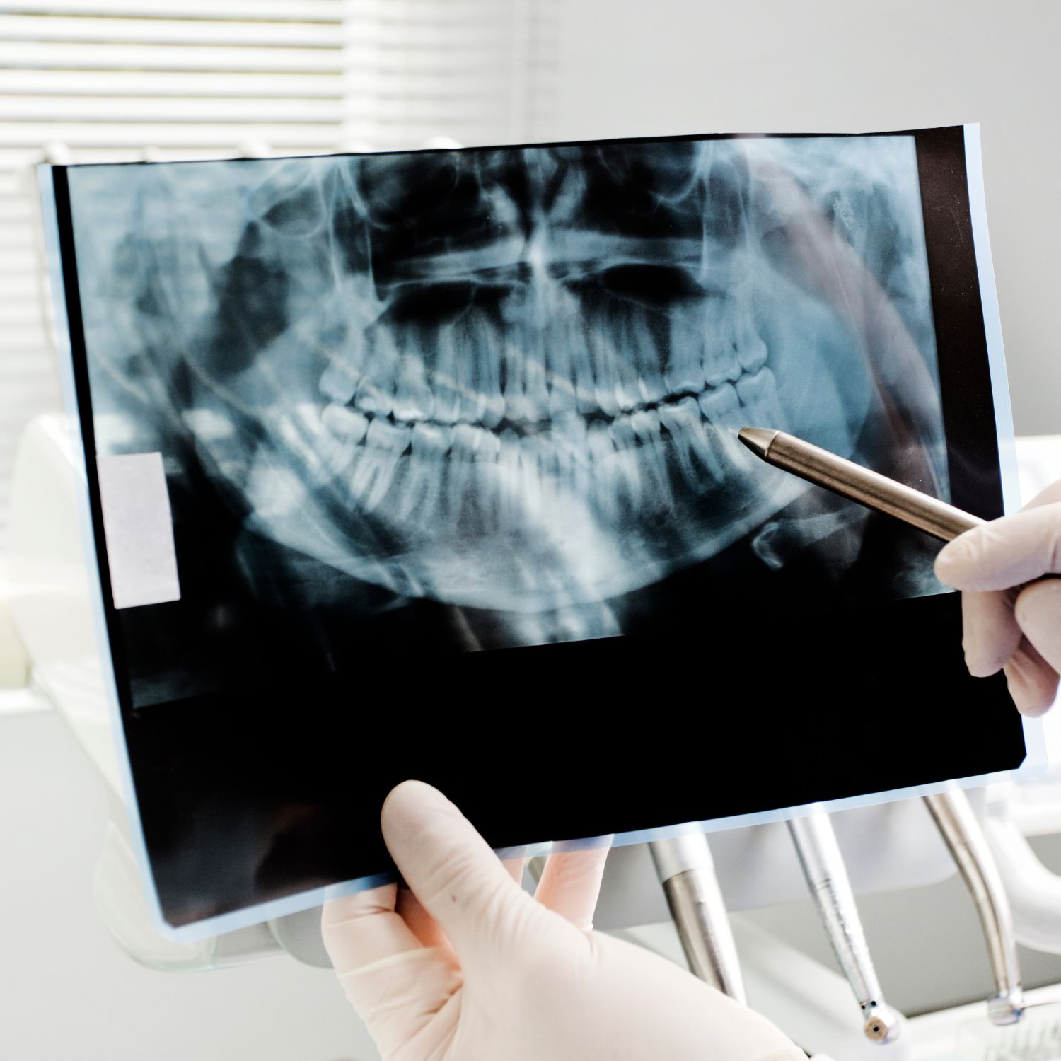
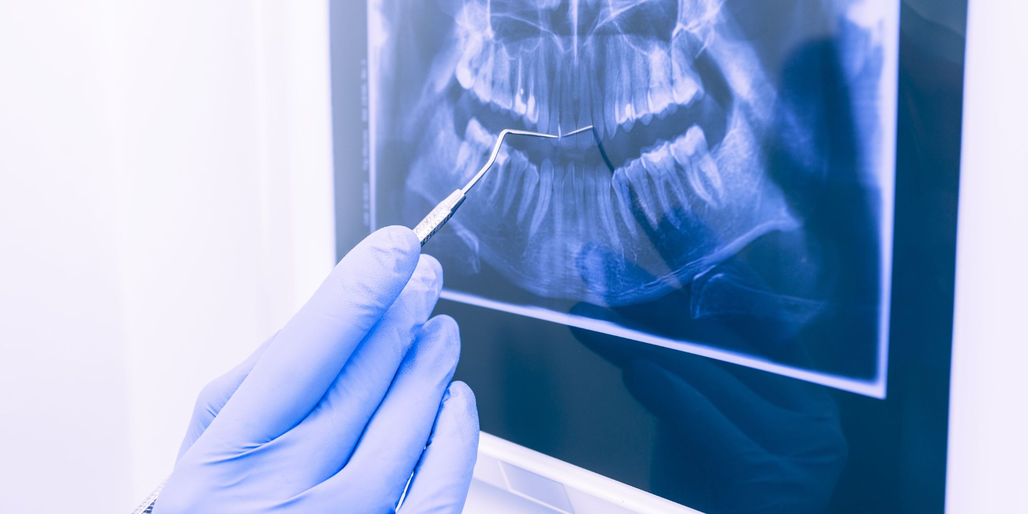
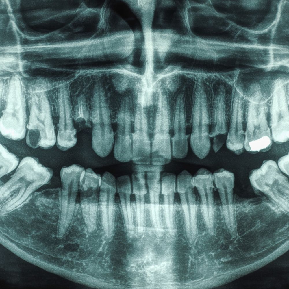
X-ray oral screening uses a form of electromagnetic radiation to take pictures of the teeth, gums and jaws. The X-ray machine emits a small amount of radiation, which passes through the oral cavity and is absorbed by various tissues to varying degrees. X-rays passing through the tissues create an image on a digital sensor or x-ray film, allowing the dentist to identify any abnormalities or potential problems.
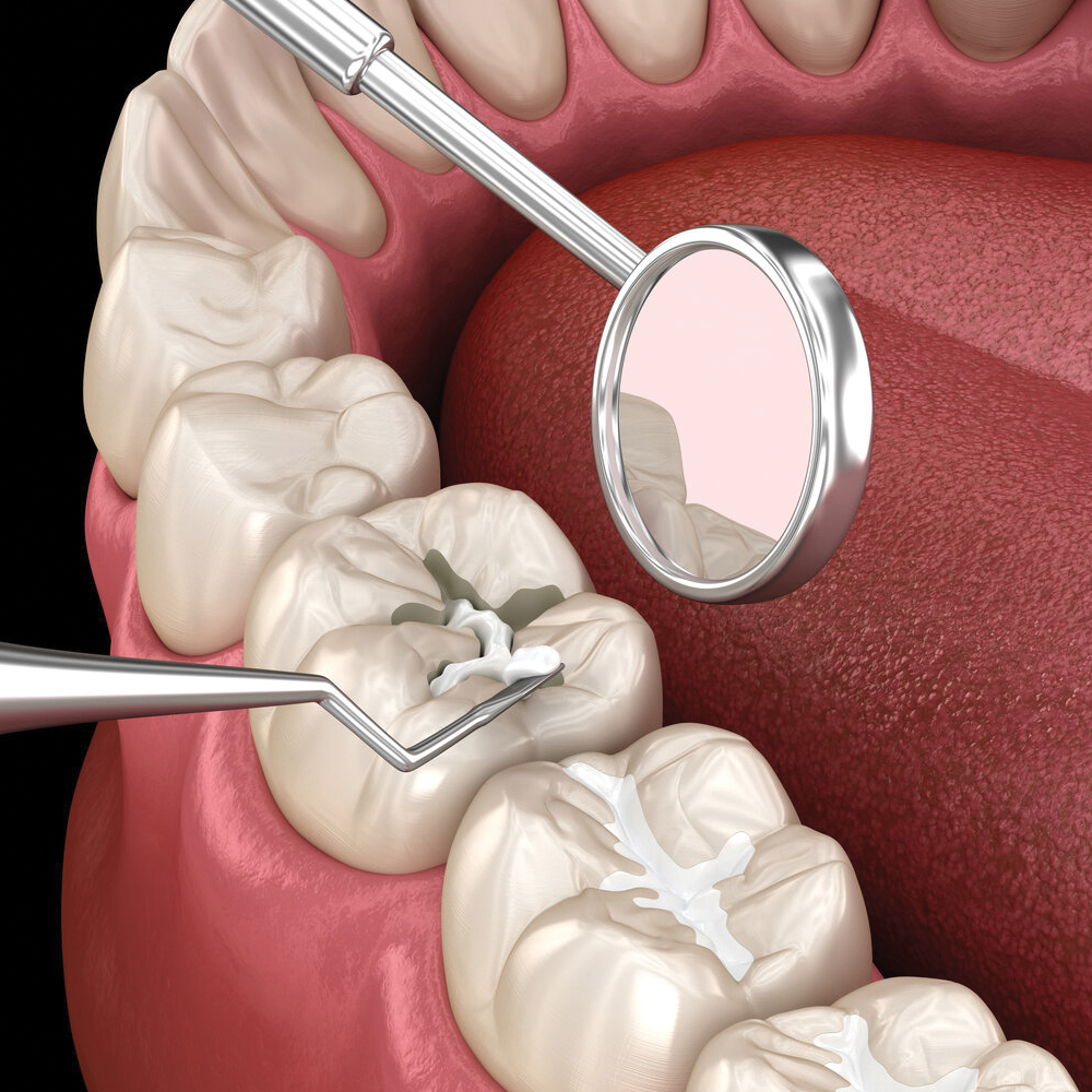
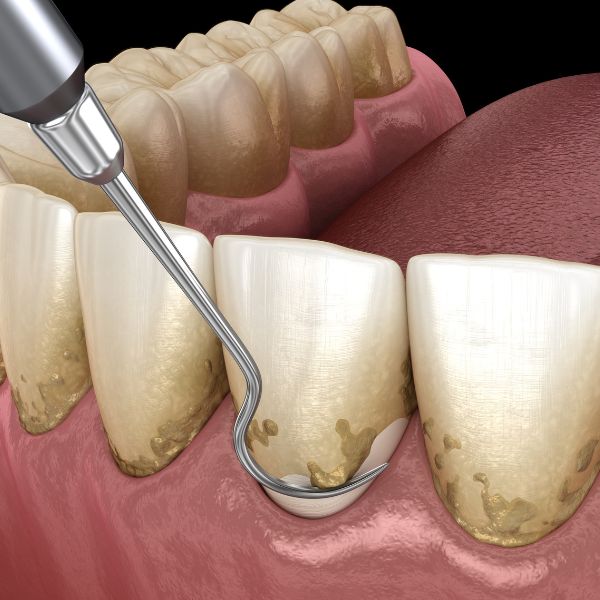
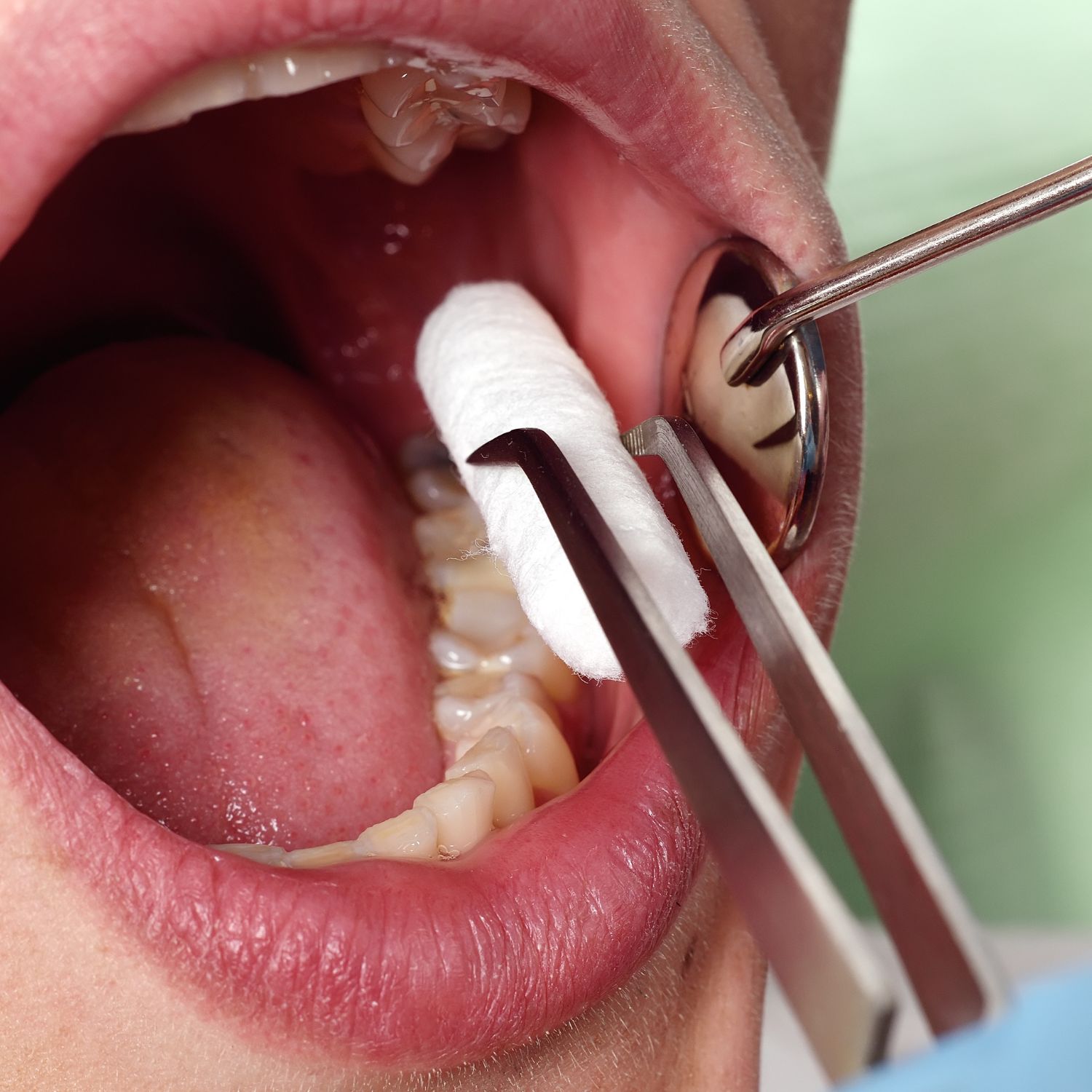
There are two basic types of X-ray oral examinations: intraoral and extraoral. Intraoral X-rays involve placing a small sensor or film inside the mouth to take detailed images of individual teeth. These images help dentists identify problems such as tooth decay, gum disease, infections, and abnormalities in tooth structure. On the other hand, extraoral radiographs capture images of the entire oral cavity, including the jaws and surrounding structures. These images are used to assess tooth development, monitor the position of wisdom teeth and identify any fractures or tumors.
X-ray screening is safe and radiation exposure is minimal. Dentists take precautions by using lead aprons and thyroid collars to protect patients from unnecessary radiation. In addition, modern X-ray machines emit lower levels of radiation compared to older models, making the procedure even safer.
Why is x-ray oral examination important?
It is essential for maintaining and improving oral health. They provide valuable information that cannot be obtained through visual examination alone. Here are some reasons why oral exams are important:
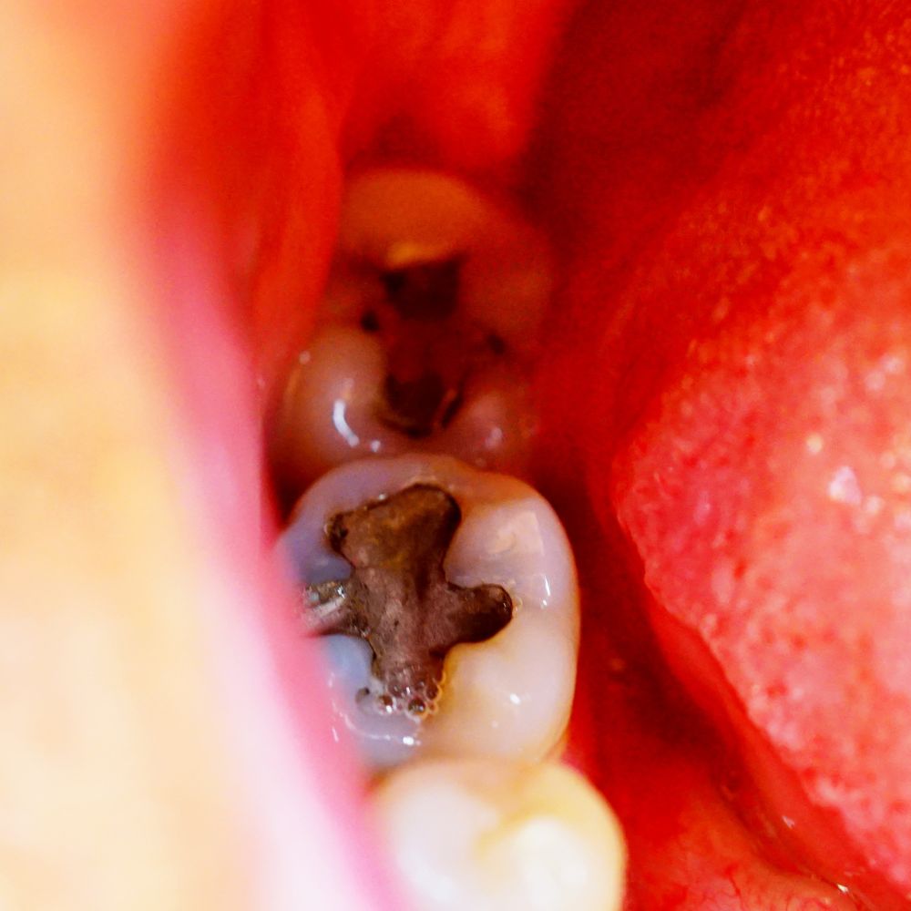
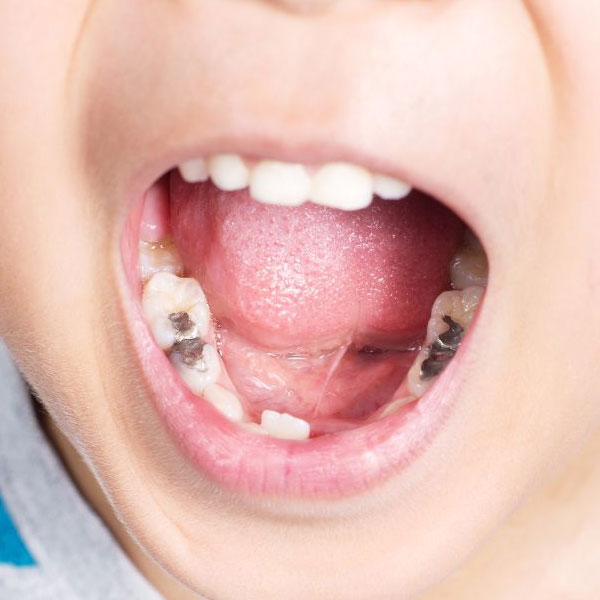
Detecting Hidden Dental Problems: Oral exams with X-rays can help identify dental problems that are not visible to the naked eye. For example, X-rays can reveal cavities between teeth, bone loss due to gum disease, impacted wisdom teeth, and infections in the jawbone. By identifying these problems early, dentists can prevent further damage and provide appropriate treatment.
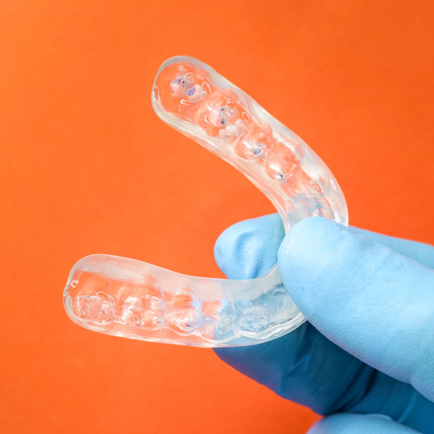
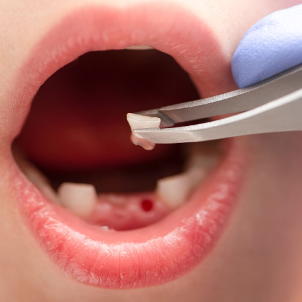
Assessment of tooth and jaw development: X-rays are particularly useful in assessing the development of the teeth and jaws, especially in children and adolescents. Dentists can determine if the teeth are erupting properly and monitor any abnormalities in the development of the jaws. This information is vital to planning orthodontic treatment and ensuring proper tooth alignment.
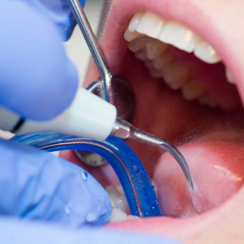
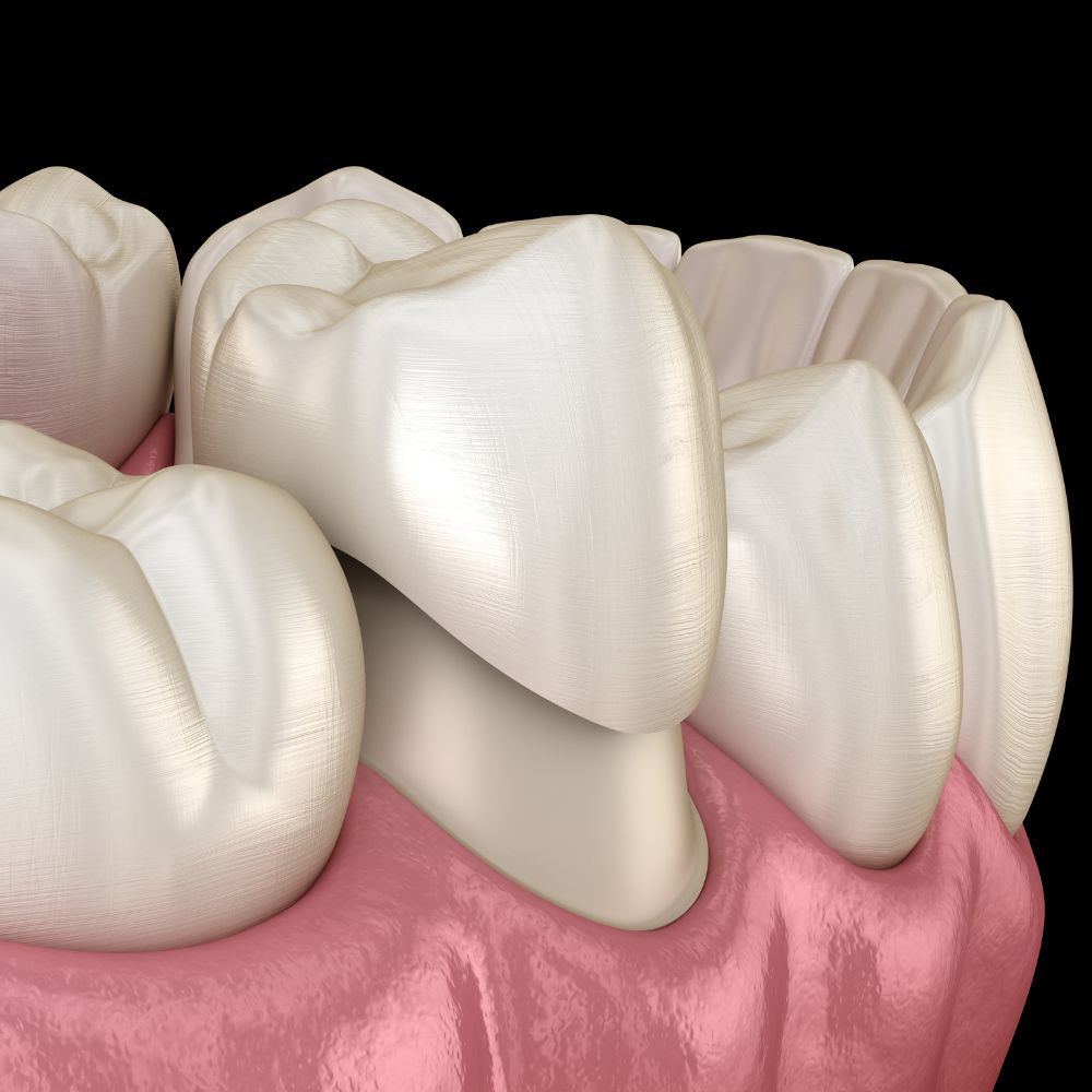
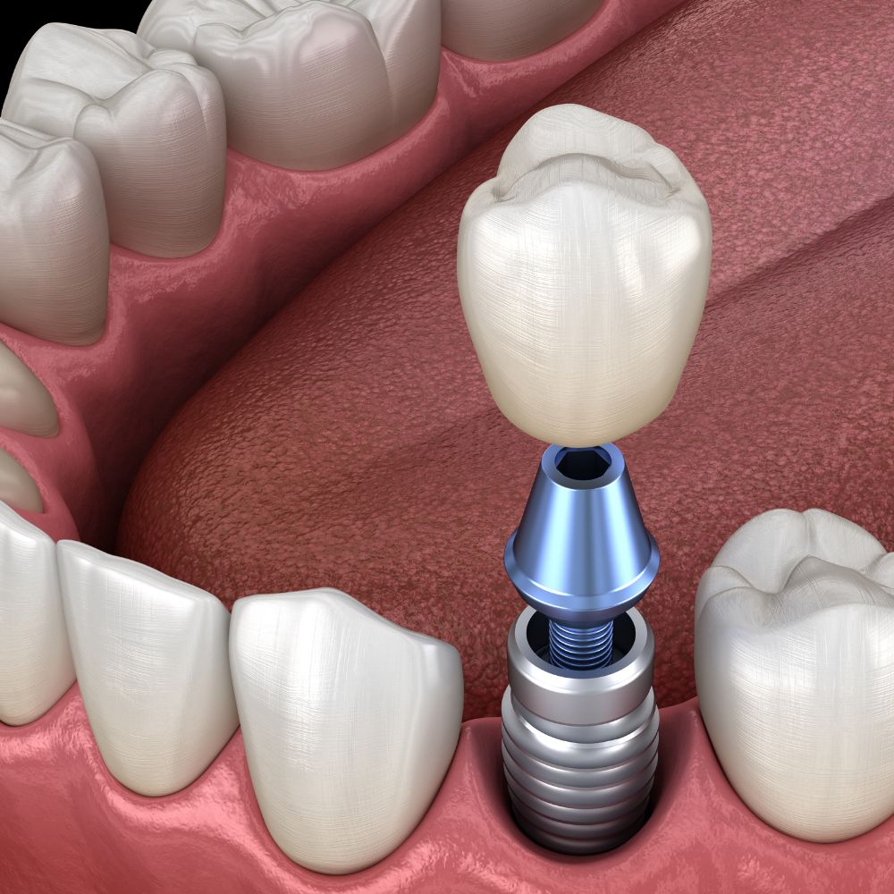
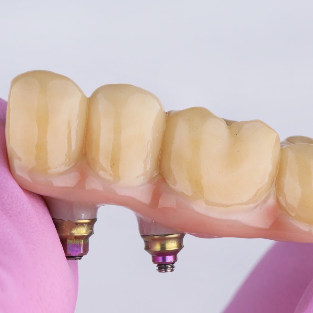
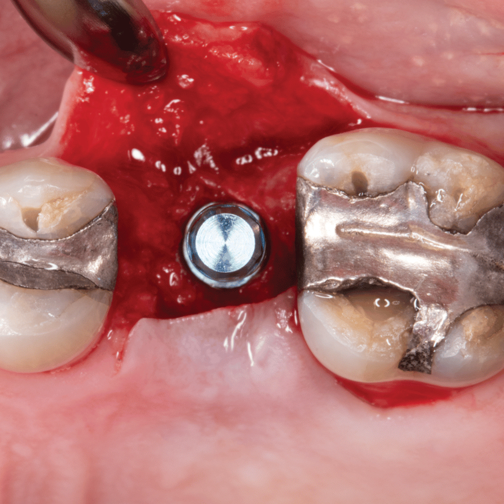
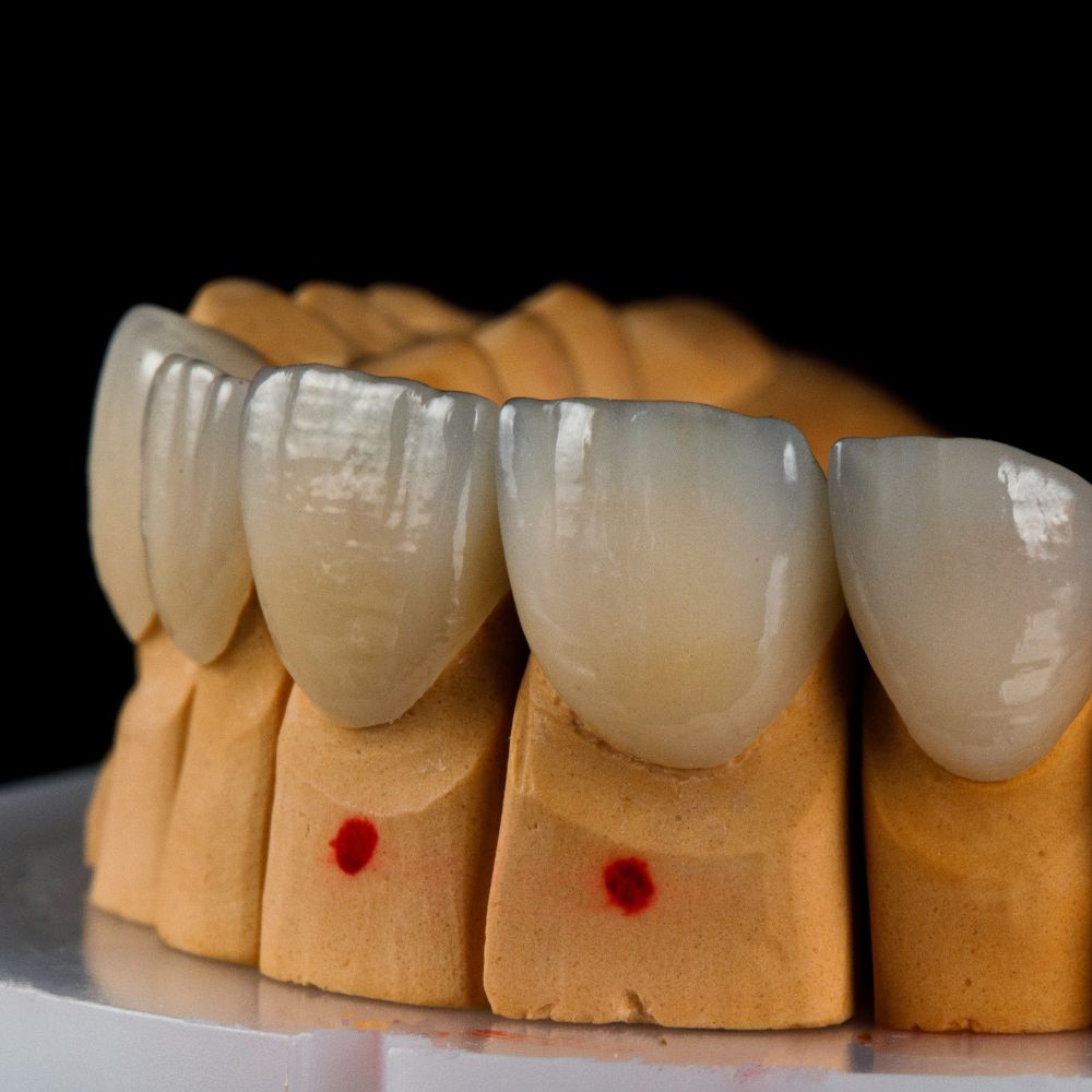
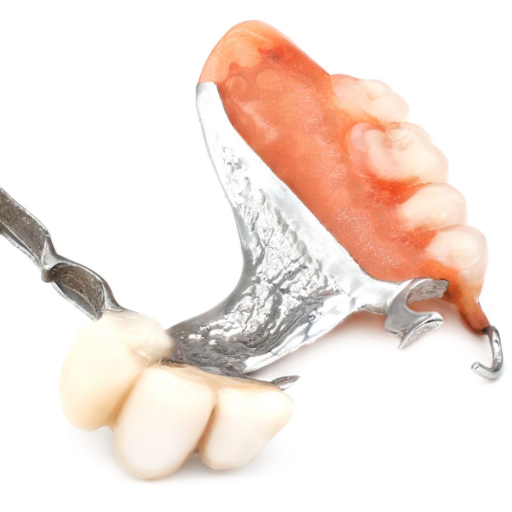
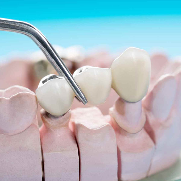
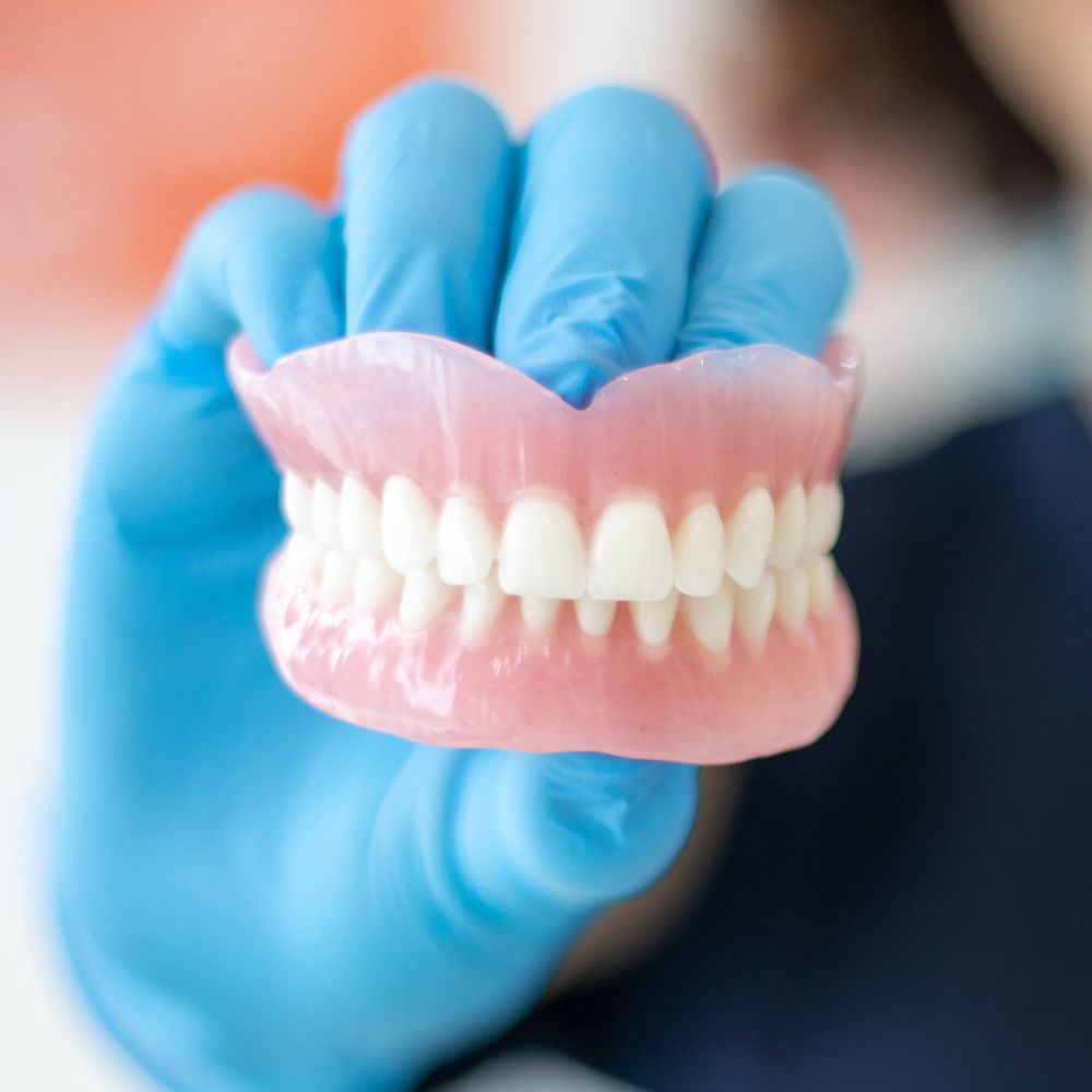
Evaluation of previous dental work: X-rays allow dentists to assess the success and longevity of previous dental treatments. They can check the integrity of fillings, crowns, bridges and implants, making sure they are working properly and not causing problems.
Diagnosis of oral diseases: X-ray oral examinations play a vital role in the diagnosis of oral diseases, such as oral cancer. X-rays can reveal abnormal growths or tumors in the oral cavity, allowing for early detection and intervention. This early diagnosis greatly improves the chances of successful treatment.